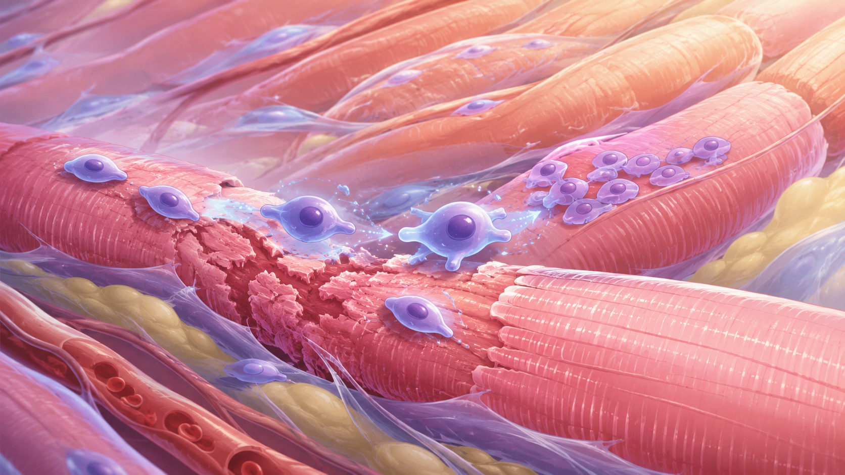
Imagine a line of molecular dominoes. The first block falls when a receptor on the cell surface is activated. That knocks down the second, which knocks down the third, and so on, each protein activating the next in a sequence that, in the end, determines whether the cell will grow, divide, survive, or enter atrophy. That line of dominoes has a name: PI3K/Akt/mTOR. And it is, without exaggeration, one of the most studied and most clinically relevant signaling cascades in all of cell biology.
IGF-1 LR3 is one of the tools researchers use to study that cascade with precision. And understanding why requires understanding both what this pathway does and why studying it with native IGF-1 has limitations that the synthetic analog overcomes.
PI3K/Akt/mTOR is the pathway that tells the cell: you have enough resources, you can grow. Understanding how to activate and modulate it is one of the most promising frontiers of applied biochemistry.
The cascade, from receptor to ribosome
It all begins at the IGF-1R receptor. When a molecule of IGF-1, or of its analog IGF-1 LR3, fits into this transmembrane receptor, it auto-phosphorylates: it activates itself. That activation recruits adaptor proteins called IRS-1 and IRS-2 (Insulin Receptor Substrate), which in turn recruit and activate PI3K, phosphatidylinositol 3-kinase.
PI3K phosphorylates lipids in the cell membrane, creating signals that activate a protein called Akt, also known as PKB. Akt is one of the central nodes of the system: it phosphorylates dozens of target proteins with effects ranging from cell survival to the control of glucose metabolism. Among its most studied targets is mTOR, mechanistic Target of Rapamycin.
| Start | IGF-1 LR3 binds the IGF-1R receptor on the cell surface |
| IGF-1R → IRS-1/IRS-2 | Phosphorylation of adaptor substrates, recruitment of PI3K |
| PI3K → PIP3 | Phosphorylation of membrane lipids, generation of the second messenger PIP3 |
| PIP3 → Akt (PKB) | Activation of Akt, central node of anabolic signaling and survival |
| Akt → mTORC1 | Activation of the mTOR complex 1, master regulator of protein synthesis |
| mTORC1 → p70S6K / 4E-BP1 | Phosphorylation of ribosomal targets, activation of protein synthesis |
| Akt → FOXO1/3 | Suppression of pro-atrophy transcription factors, inhibition of MuRF1 and MAFbx |
| Akt → GSK3β | Inhibition of GSK3β, activation of glycogen synthesis |
mTOR, the node science wants to understand
mTOR is currently one of the most studied proteins in the entire field of cell biology. It is no exaggeration: there are more than 150,000 publications in PubMed mentioning mTOR, and the pace of research continues to accelerate. The reason is that mTOR functions as a sensor of cellular resources, it integrates signals of nutrient availability, energy, growth factors, and stress. When resources are available and the anabolic signals arrive (as via IGF-1R), mTOR signals to the cell: you can grow.
When mTOR is activated via Akt, it phosphorylates two key proteins: p70S6K, which activates the ribosomal machinery of protein synthesis, and 4E-BP1, which releases the translation-initiation factor eIF4E. The result is an increase in protein synthesis, especially of proteins associated with growth and cell proliferation. In muscle cells, that process is directly related to the growth of muscle fibers.
Why research mTOR with IGF-1 LR3? Native IGF-1, when used experimentally, is sequestered by the IGFBPs, which reduces its capacity to activate the receptor and the downstream cascade consistently. IGF-1 LR3, with affinity ~1000x lower for the IGFBPs, allows the IGF-1R/PI3K/Akt/mTOR pathway to be activated more predictably and sustainedly, making experiments more interpretable.
The dual pathway, MAPK and the proliferation-differentiation balance
The PI3K/Akt/mTOR cascade is not the only route activated by IGF-1R. In parallel, the receptor triggers the MAPK pathway, Mitogen-Activated Protein Kinase, through the Ras/Raf/MEK/ERK sequence. While the PI3K/Akt pathway is more associated with anabolism and cell survival, the MAPK pathway is strongly linked to proliferation and differentiation.
In muscle cells, this creates an interesting research question: depending on the balance between these two cascades, the cell can be directed to grow (hypertrophy), divide (satellite-cell proliferation), or differentiate into mature myofibers. Understanding how IGF-1 LR3 modulates that balance, and whether there are experimental parameters (concentration, exposure time, cell state) that influence which pathway predominates, is exactly the kind of question this line of research is investigating.
Two cascades, one receptor, multiple possible cell fates. The balance between PI3K/Akt and MAPK is one of the most fascinating frontiers of cell-growth biology.
IGF-1 LR3 as a tool, and what that means for research
In this context, the value of IGF-1 LR3 as a research tool becomes evident. To study the PI3K/Akt/mTOR pathway in response to IGF-1R activation, the researcher needs a molecule that activates the receptor consistently, sustainedly, and without the interference of endogenous binding proteins. IGF-1 LR3, with an estimated half-life of 20 to 30 hours in animal models and minimal affinity for the IGFBPs, offers exactly that profile.
In vitro studies with muscle cell lines document the activation of the IGF-1R/IRS-1/PI3K/Akt cascade in response to IGF-1 LR3. Studies with bovine satellite cells published in 2024 investigate how IGF-1 regulates myogenic differentiation via PI3K/AKT, a line of research that, if the data are confirmed in translational models, may shed light on fundamental mechanisms of muscle growth and regeneration. The convergence between this signaling axis and questions such as sarcopenia, cellular aging, and muscle biology represents one of the most productive research directions available today.
PI3K, Akt, mTOR. Three letters each. But what these proteins do, and how to understand them better, may be part of one of the most important stories molecular biology is writing right now.